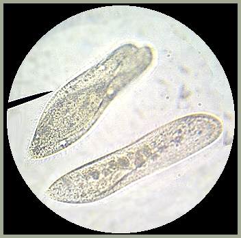
The upper image shows two Paramecia reproducing by conjugation. The two are joined and a cytoplasmic bridge is formed between the two animals. The nuclei from both Paramecia divide several times. One of the divided nuclei from each Paramecium travels across the bridge and fuses with one of the nuclei from the other animal. The nuclei form another nucleus and cell, the others dissolve, and the new cell separates. This cell can divide several times, creating a wholly new Paramecium in each case.
The lower image shows a single Paramecium. Paramecia travel rather quickly, and they "corkscrew" as they move through the water. These creatures were slowed (but not stopped) by adding methyl cellulose to the water sample and it enabled me to obtain reasonably clear images at 400x. The oral groove, which unmistakably identifies the Paramecium is not visible in either image.
Back To Image Index Page

